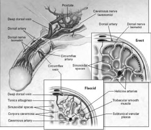
A normal erection requires the penis’ nerves and blood vessel systems to be intact and to have appropriate hormonal levels, but also is moderated by psychological factors. The penis is stimulated by both the autonomic nervous system — the part of the nervous system that functions independent of our conscious thought — and the somatic nervous system — the nervous system responsible for sensation and contraction of muscles attached to the penis. The glans or head and body of the penis have numerous sensory nerve endings that send messages of pain, temperature and touch back to the brain. The motor nerves stimulate the muscles in the pelvis and penis — the ischiocavernosus and bubocavernosus muscles — that are necessary to produce a rigid erection and ejaculation. The autonomic nervous system stimulates the rectum, bladder, prostate and sphincters, includes the cavernous nerve that stimulates the penis and controls the flow of blood during and after erection.
With sexual stimulation, the cavernous nerves release chemicals that significantly increase blood flow to the penis. The erectile tissue of the penis rapidly fills, expands and becomes erect. During sexual activity, the bulbocavernous and ischiocavernous muscles of the penis are stimulated, which compresses the base of the penis to make the penis even harder.
During emission, seminal fluid is released from the seminal vesicles and the prostate into the urethra. The bladder sphincter then closes, and the seminal fluid becomes trapped. As the amount of fluid builds in the urethra, the pressure increases and the sensation of the inevitability of ejaculation is experienced. The bulbocavernous muscle contracts and expels the semen forcibly from the urethra. Orgasm normally coincides with ejaculation.
Detumescence, or loss of erection, occurs shortly thereafter and is produced by the breakdown of the factors that cause erection.
Anatomy and Mechanism of Penile Erection
The cavernous nerves travel from the underside of the penis to the prostate. These nerves regulate blood flow within the penis during erection and flaccidity. In the flaccid state, inflow through the arteries is minimal and there is free outflow via the small veins exiting the spongy tissue just under the thick tunica (thick membrane surrounding the spongy tissue). During erection, the smooth muscle in the penis relaxes while the arteries widen to pump in more blood that expands the three cavities of the penis — also called sinusoidal spaces — to lengthen and enlarge the penis. The expansion of these cylinders compresses the small veins and reduces the outflow of blood.
Future Directions
Innovative research over the past several years has resulted in significant strides and improvement to understanding the anatomy and physiology of sexual function. For instance, increasing knowledge about details of the cavernous nerves in the pelvis led to refinement of nerve-sparing prostatectomy. Understanding the biochemistry of normal sexual functioning led to the development of medications including Sildenafil, Cialis and Levitra.
Current research is focusing on further understanding of the specific physiologic pathways responsible for normal sexual function, developing new, more effective agents for managing impotence and understanding how cavernous nerves heal and what factors can hasten the healing process. Use of “gene” or “stem cell” technology may be possible in the future, allowing men and their partners to enjoy better sexual health.
